Homeostasis and Physiological Regulation for the ESAT
Updated July 2026
Homeostasis is the maintenance of a constant internal environment, essential for the health of multicellular animals. It relies on negative feedback loops to regulate variables such as blood glucose, body temperature, and water content. Understanding these systems, including the roles of insulin, glucagon, and ADH, is vital for the Biology section of the ESAT.
Homeostasis is the maintenance of internal conditions within set limits to ensure a stable environment for cellular function. This is primarily achieved through negative feedback, where a change in a variable triggers a response that counteracts and reverses the original deviation.
Understanding Homeostasis
Homeostasis is defined as the maintenance of a constant internal environment. For multicellular animals, it is vital to keep internal conditions within set limits to remain in a healthy state. This involves regulating the composition of tissue fluid, including water content, nutrient concentrations (such as glucose), and core body temperature. If these factors fluctuate significantly without being corrected, it can lead to organ failure or death.
The Mechanism of Negative Feedback
Homeostatic control relies on negative feedback, a system involving detectors and effectors. When an internal condition deviates from the normal level, the following sequence occurs:
- Detection: A sensor or detector identifies the change.
- Information Transfer: The sensor sends information to an effector.
- Response: The effector is stimulated to produce a response that reverses the initial change.
- Negative Feedback: Once the level returns to normal, the sensor detects this and signals the effector to switch off.
For example, if core body temperature rises, the thermoregulatory centre in the brain detects the change and triggers cooling mechanisms. Once the temperature drops back to normal, those cooling mechanisms are deactivated to prevent overcooling.
Regulation of Blood Glucose Levels
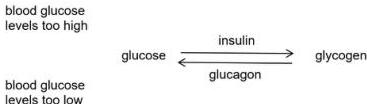
The liver and pancreas work together to control the concentration of glucose in the blood. This is essential because extreme fluctuations can lead to a loss of consciousness or death. Control is achieved through two primary hormones: insulin and glucagon.
Responding to High Blood Glucose
After consuming a meal rich in carbohydrates, blood glucose levels rise as glucose is absorbed into the bloodstream. The islet cells in the pancreas detect this increase and secrete insulin into the blood plasma. Insulin travels to the liver, where it stimulates liver cells to take up glucose and convert it into the storage carbohydrate glycogen. Insulin also encourages other body cells to absorb more glucose for use in aerobic respiration.
Responding to Low Blood Glucose
If glucose levels fall, such as during exercise or long periods without food, the islet cells of the pancreas detect the decrease and release glucagon. Glucagon acts on the liver cells, causing them to convert stored glycogen back into glucose, which is then released into the blood to raise the concentration back to normal.
Worked Example: Insulin Concentrations
Question: In a healthy individual, plasma insulin levels are approximately before and after a meal, but rise to during a meal. Why does this happen, and why must insulin be injected rather than swallowed as a tablet?
Step 1: Explaining the rise: During a meal, carbohydrates are digested into glucose and absorbed. The rise in blood glucose is detected by the pancreatic islet cells, which secrete insulin to stimulate the conversion of glucose to glycogen in the liver. Once levels return to normal, secretion stops.
Step 2: Explaining the injection: Insulin is a protein. If taken orally, it would enter the stomach where protease enzymes would digest it into amino acids. This would destroy its molecular structure, rendering it unable to function as a hormone. Therefore, it must be injected directly into the bloodstream.
Diabetes Mellitus
Diabetes occurs when the body's ability to regulate blood glucose is impaired.
- Type 1 Diabetes: Usually appears in young people and is often an autoimmune disease where the immune system attacks the pancreatic islet cells. This results in an inability to produce sufficient insulin. Symptoms include fatigue, extreme thirst, weight loss, and frequent urination. It is managed with a regulated diet, frequent blood testing, and regular insulin injections.
- Type 2 Diabetes: The most common form, often linked to obesity, poor diet, and lack of exercise. It involves insulin resistance, where body cells no longer respond effectively to insulin. It can often be managed through lifestyle changes, though it may be influenced by heredity.
Regulation of Water Content (Osmoregulation)
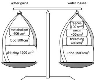
The body must maintain a balance between water gain (from drinking, eating, and metabolic water from respiration) and water loss (via sweat, exhaled air, urine, and faeces).
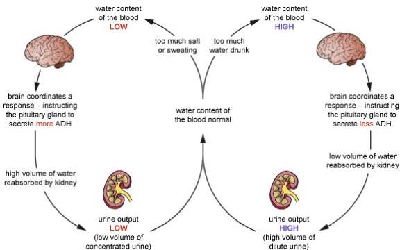
The brain monitors blood water levels and signals the pituitary gland to adjust the secretion of ADH (antidiuretic hormone). ADH travels in the blood to the kidneys, specifically acting on the collecting ducts of the nephrons.
- High Water Content (e.g., a cold day): The brain detects high water levels and signals the pituitary to release less ADH. The collecting ducts become less permeable, less water is reabsorbed into the blood, and the kidneys produce a large volume of dilute urine.
- Low Water Content (e.g., a hot day/exercise): The brain signals the pituitary to release more ADH. This makes the collecting ducts more permeable. Water leaves the ducts by osmosis and re-enters the blood capillaries. The result is a small volume of concentrated urine.
Regulation of Body Temperature
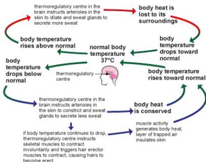
Thermoregulation is controlled by the thermoregulatory centre in the brain, which monitors the temperature of the blood and receives data from skin receptors.
Cooling Mechanisms (Response to heat)
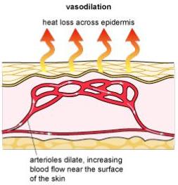
- Vasodilation: Arterioles (not capillaries) near the skin surface dilate. This allows more blood to flow through capillaries close to the skin, increasing heat loss by radiation.
- Sweating: Sweat glands secrete sweat onto the skin surface. As the water in the sweat evaporates, it takes heat energy from the skin, cooling the body.
Warming Mechanisms (Response to cold)
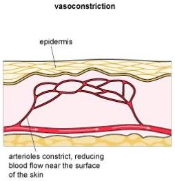
- Vasoconstriction: Arterioles near the skin surface constrict, reducing blood flow to the surface and conserving heat.
- Shivering: Rapid muscle contractions generate heat through increased respiration.
- Erection of hairs: Small muscles pull hairs upright to trap an insulating layer of air against the skin.
Key takeaways
- Homeostasis is the maintenance of a constant internal environment via negative feedback loops.
- Insulin lowers blood glucose by stimulating the liver to convert glucose to glycogen; glucagon raises it by doing the opposite.
- Type 1 diabetes is an autoimmune lack of insulin; Type 2 is typically a lifestyle-linked resistance to insulin.
- ADH regulates water reabsorption in the kidney by increasing the permeability of the collecting ducts.
- Thermoregulation involves vasodilation and sweating to lose heat, or vasoconstriction and shivering to conserve it.
For ESAT questions, remember that negative feedback always works to reverse a change. If a question describes a hormone increasing a variable, it is likely part of a feedback loop intended to counteract an initial decrease.
Do not confuse the roles of insulin and glucagon. Insulin 'tucks' glucose away (lowers blood levels), while glucagon makes glucose 'gone' from storage (raises blood levels). Also, ensure you specify that insulin acts on the liver to produce glycogen.
Homeostasis illustrates the principle of 'dynamic equilibrium'. The internal environment is not perfectly static but fluctuates slightly around a set point; negative feedback ensures these fluctuations remain within a safe, narrow range.
Frequently asked questions
What is the difference between a detector and an effector?
A detector (or sensor) is a structure, such as the thermoregulatory centre in the brain or islet cells in the pancreas, that monitors a specific variable. An effector is a muscle or gland, such as a sweat gland or the liver, that carries out a response to restore the variable to its set point.
How does ADH actually change urine concentration?
ADH makes the walls of the collecting ducts in the kidney nephrons more permeable to water. This allows more water to be reabsorbed back into the blood by osmosis, leaving behind a smaller, more concentrated volume of urine.
Why are arterioles mentioned instead of capillaries in thermoregulation?
Arterioles have muscle in their walls that can contract or relax to change the diameter of the vessel (vasoconstriction or vasodilation). Capillaries do not have muscle in their walls and therefore cannot change their diameter to regulate blood flow in this way.
What are the main symptoms of Type 1 diabetes?
Common symptoms include extreme thirst, frequent urination (polyuria), fatigue, weight loss, and the presence of glucose in the urine. These occur because the body cannot effectively manage the high concentration of glucose in the blood.