Animal Physiology for the ESAT
Updated July 2026
An in depth guide to human organ systems including the respiratory, circulatory, digestive, and excretory systems. You will learn the mechanics of ventilation, the structure of the heart, blood composition, and homeostatic regulation of glucose and water. Understanding these systems is vital for interpreting biological data on the ESAT.
Multicellular organisms require specialized organ systems and exchange surfaces to overcome the limitations of a small surface area to volume ratio, ensuring efficient transport of nutrients, gases, and waste products through processes like diffusion, active transport, and negative feedback.
The Respiratory System
The respiratory system is located within the thorax, the chest cavity. Air enters through the nose and mouth, passing through the larynx and into the trachea. The trachea is kept open by rings of cartilage. It bifurcates into two bronchi, which lead into the lungs and branch further into bronchioles. At the end of these bronchioles are the alveoli, where gas exchange occurs.
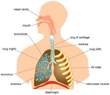
The thorax is protected by the rib cage, which features intercostal muscles. Beneath the lungs lies the diaphragm, a muscular sheet. Together, these structures facilitate ventilation. The primary function is to supply oxygen for aerobic respiration and remove the waste product, carbon dioxide.
Inhaled versus Exhaled Air
Air undergoes changes as it moves through the body. Inhaled air contains more oxygen and less carbon dioxide and water vapour than exhaled air. It also contains particles like dust and pathogens. The bronchi are lined with cells that produce mucus to trap these particles: cilia then sweep the mucus up to the trachea to be swallowed and destroyed by stomach acid.
Worked Example: Exercise 40 What is the correct order for a molecule of carbon dioxide being exhaled? The correct sequence follows the reverse path of inhalation: alveoli to bronchioles, then bronchi, then trachea, and finally the nose or mouth.
Worked Example: Exercise 41 Tar in cigarettes paralyses cilia. This results in the smoker being more likely to develop lung infections because the mucus containing trapped pathogens is not cleared effectively.
Ventilation: The Mechanics of Breathing
Ventilation is the exchange of air between the lungs and the atmosphere. In vertebrates, this is driven by volume and pressure changes in the thorax.
- Inhaling (Breathing In): The intercostal muscles contract, pulling the ribs up and out. Simultaneously, the diaphragm contracts and flattens. This increases the volume of the thoracic cavity, lowering the internal air pressure. Air rushes in to equalise the pressure.
- Exhaling (Breathing Out): This is usually passive. The intercostal muscles relax, ribs move down and in, and the diaphragm relaxes into a dome shape. This decreases the volume, increases pressure, and forces air out.
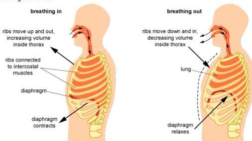
Worked Example: Exercise 42 Inhaling involves the ribs moving up and out and the thoracic pressure decreasing. Therefore, statements 1 and 3 are correct.
Worked Example: Exercise 43 A logical sequence for exhalation is: the ribs move down and inwards (4), the volume inside the thorax decreases (6), and air passes through the bronchi (5) and trachea (3).
Gas Exchange and the Alveoli
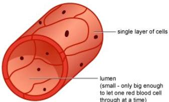
Gas exchange occurs via diffusion across the walls of the alveoli and capillaries, both of which are only one cell thick to minimise diffusion distance. Oxygen diffuses from the alveoli into the blood, while carbon dioxide diffuses from the blood into the alveoli.
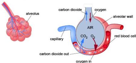
Adaptations for efficient exchange include:
- Large surface area provided by millions of alveoli.
- Short diffusion pathways (single layer of cells).
- Constant blood flow to maintain steep concentration gradients.
Worked Example: Exercise 44 Increasing the heart rate maintains the concentration gradient of gases more effectively, thereby increasing the rate of diffusion. Tar or a reduction in alveolar surface area would decrease the rate.
Surface Area to Volume Ratio (SA:V)
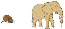
SA:V ratio describes how much surface area is available relative to the volume of an organism. Small organisms, like bacteria, have a large SA:V ratio and can rely on simple diffusion. Large organisms have a small SA:V ratio and require specialised transport systems.
Calculation Example: 3 mm Cube
- Surface Area () = .
- Volume () = .
- Ratio = .
Worked Example: Exercise 46 For a cube with cm sides:
- .
- .
- .
- The ratio is .
The Circulatory System
The circulatory system consists of the heart and a network of vessels: arteries, veins, and capillaries. Specific organs have named vessels: Coronary (heart), Pulmonary (lungs), Hepatic (liver), and Renal (kidneys).
Blood Vessels
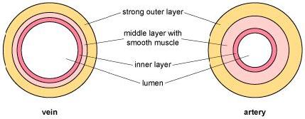
- Arteries: Carry blood away from the heart at high pressure. They have thick, muscular, and elastic walls and a narrow lumen. They generally carry oxygenated blood (except the pulmonary artery).
- Veins: Return blood to the heart at lower pressure. They have thinner walls, a wide lumen, and valves to prevent backflow. They generally carry deoxygenated blood (except the pulmonary vein).
- Capillaries: Tiny vessels with walls one cell thick, allowing for the exchange of substances between blood and tissues.
The Heart and ECGs
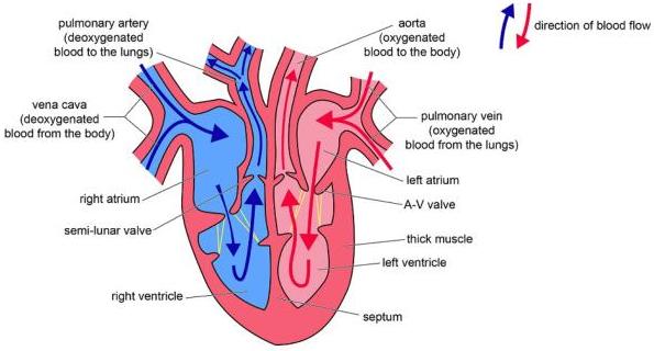
The heart is a muscular pump. Deoxygenated blood enters the right side and is pumped to the lungs. Oxygenated blood returns to the left side and is pumped to the rest of the body.
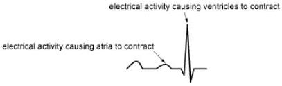
Heart activity is coordinated by electrical impulses, which can be recorded using an Electrocardiogram (ECG). Adrenaline increases the heart rate, which is visible on an ECG as more frequent cycles.
Worked Example: Exercise 47 Oxygen levels (partial pressure, ) vary. The vena cava (returning from body) has a of mmHg. The pulmonary vein (returning from lungs) is mmHg. The aorta and renal artery (systemic circulation) have a of mmHg. Statements a and f are true.
Worked Example: Exercise 49 Adrenaline increases the heart rate, so Graph B, which shows more beats in the same time interval, is the correct representation.
Composition of the Blood
Blood consists of cells suspended in plasma (which makes up percent of the volume).
- Plasma: A watery liquid transporting dissolved glucose, urea, amino acids, hormones, antibodies, and carbon dioxide. It also distributes heat.
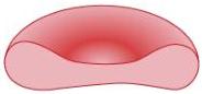
- Red Blood Cells: Biconcave discs with no nucleus, containing haemoglobin to transport oxygen.
- White Blood Cells: Phagocytes engulf and digest pathogens: lymphocytes produce antibodies to target antigens.
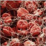
- Platelets: Cell fragments involved in clotting. They help convert soluble fibrinogen into insoluble fibrin, creating a mesh to trap cells and form a scab.
The ABO Blood Group System
Blood groups are determined by proteins on the red blood cell surface. The and alleles are co-dominant, while is recessive. Antibodies in the plasma react against foreign antigens.
- Group A: Has A antigens and anti-b antibodies. Can receive from A or O.
- Group B: Has B antigens and anti-a antibodies. Can receive from B or O.
- Group AB: Has A and B antigens, no antibodies. Universal recipient.
- Group O: No antigens, has anti-a and anti-b antibodies. Universal donor but can only receive O.
The Digestive System
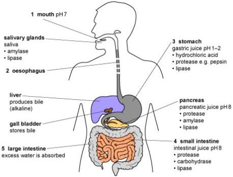
Digestion is the breakdown of large, insoluble molecules into small, soluble ones for absorption. The path is: mouth to oesophagus to stomach to small intestine to large intestine.
Peristalsis and Digestion
Food is moved by peristalsis, waves of muscular contraction. Digestion can be mechanical (teeth, stomach churning) or chemical (enzymes).
- Proteins: Digested by proteases (e.g., pepsin in the stomach at low pH: pancreatic proteases in the small intestine at alkaline pH) into amino acids.
- Carbohydrates: Starch is broken down by amylase (salivary and pancreatic) into smaller chains and then monosaccharides like glucose.
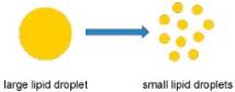
- Lipids: Emulsified by bile (made in liver, stored in gall bladder) to increase surface area, then broken down by lipases into fatty acids and glycerol.
Absorption in the Small Intestine
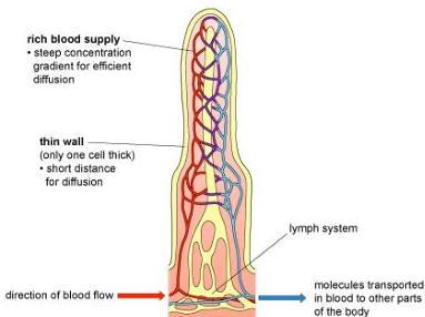
The small intestine wall features villi, which increase surface area. Nutrients are absorbed via diffusion and active transport into the blood or lymph (for fats). Water is absorbed in both the small and large intestines. Indigestible waste (fibre) is egested as faeces.
The Excretory System and Homeostasis
Excretion is the removal of toxic waste products. Key organs include the lungs (CO2), liver (deamination of amino acids to urea), and kidneys.
The Kidneys and the Nephron
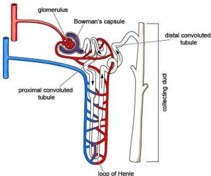
Kidneys filter blood to remove urea and adjust ion and water content. Each kidney contains millions of nephrons.
- Ultrafiltration: High pressure in the glomerulus forces water, ions, glucose, and urea into the Bowman's capsule. Large proteins and blood cells remain in the capillary.
- Selective Reabsorption: As filtrate moves through the tubule, glucose is reabsorbed via active transport. Water is reabsorbed by osmosis.
- Excretion: Remaining waste moves to the collecting duct as urine, then to the ureter, bladder, and out the urethra.
Homeostasis and Negative Feedback
Homeostasis maintains a constant internal environment via negative feedback loops. If a level deviates, a detector triggers an effector to reverse the change.
- Blood Glucose: Controlled by the pancreas. High glucose triggers insulin (liver converts glucose to glycogen). Low glucose triggers glucagon (liver converts glycogen to glucose).
- Water (Osmoregulation): Controlled by ADH from the pituitary gland. High ADH makes the nephron collecting ducts more permeable, reabsorbing more water into the blood, producing concentrated urine.
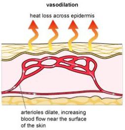
- Temperature: Monitored by the brain. A rise in temperature causes vasodilation (arterioles dilate to increase blood flow to the skin) and sweating. A fall causes vasoconstriction, shivering, and hair erection.
Key takeaways
- Ventilation is driven by pressure changes caused by the contraction and relaxation of the diaphragm and intercostal muscles.
- Arteries have thick walls and narrow lumens to carry high pressure blood, while veins have valves and wide lumens for low pressure blood.
- Digestion involves mechanical breakdown and chemical enzymes, with absorption occurring primarily in the small intestine via villi.
- The nephron performs ultrafiltration of the blood and selective reabsorption of essential nutrients like glucose and water.
- Homeostasis uses negative feedback and hormones like insulin, glucagon, and ADH to maintain internal stability.
When answering questions on the circulatory system, remember that 'Arteries' move 'Away' from the heart and 'Veins' go 'In' to the heart. Be careful with pulmonary vessels, as they are the exceptions to the oxygenation rule.
Do not confuse the 'ureter', which carries urine from the kidney to the bladder, with the 'urethra', which carries urine out of the body. This is a very common point of confusion in exams.
Notice how many systems rely on maintaining a concentration gradient for diffusion. In the lungs, constant ventilation and blood flow ensure gradients are steep: in the small intestine, a rich blood supply in the villi does the same for nutrient absorption.
Frequently asked questions
How does the surface area to volume ratio change as an organism grows?
As an organism increases in size, its volume increases much faster than its surface area. This results in a smaller surface area to volume ratio, making simple diffusion insufficient for the organism's needs.
What is the difference between type 1 and type 2 diabetes?
Type 1 diabetes is an autoimmune condition where the pancreas fails to produce enough insulin. Type 2 diabetes occurs when the body's cells become resistant to insulin, often linked to lifestyle factors like obesity.
Why is the left ventricle wall thicker than the right?
The left ventricle must pump blood at a much higher pressure to reach the entire body (systemic circulation), whereas the right ventricle only pumps blood to the nearby lungs (pulmonary circulation).
What is the role of ADH in the kidneys?
ADH (antidiuretic hormone) increases the permeability of the collecting ducts in the nephrons. This allows more water to be reabsorbed into the blood, resulting in a lower volume of more concentrated urine.